
Pathology:
The heart receives oxygenated blood from lungs in its left side chambers, first the Upper Chamber (Left Atrium) and then into Lower Chamber (Left Ventricle). Situated in between Left Atrium and Left Ventricle, the Mitral Valve regulates flow of blood in one direction. The blood is then ejected into the Aorta from the Left Ventricle through the Aortic Valve. The Aorta is main blood vessel (Artery) that carries blood from heart to the rest of the body. However, in certain diseases (such as Rheumatic Valvular Heart Disease), both the Aortic & Mitral Valves malfunction either blocking blood from flowing across the valves (Aortic or Mitral Stenosis) or letting it flow back across the valve (Aortic or Mitral Regurgitation).
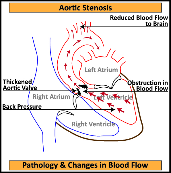
- Stenosis: In certain diseases, leaflets of Aortic and/or Mitral Valve thicken and fuse leading to reduced mobility and narrowing of the opening, reducing flow of blood from heart to body.
- Aortic Stenosis: The arteries supplying blood to the heart (Coronary Arteries) connect to the Aorta immediately after the Aortic Valve. A reduced flow of blood through these Coronary Arteries can cause chest pain (Angina on Exertion). The ascending Aorta further connects to three arteries which supply blood to both the hands and the brain. Reduction of blood flowing into the brain causes fainting or passing out (Syncope). Increase in back-pressure due to obstruction to flow of blood in the heart and lungs causes Breathlessness.
- Mitral Stenosis: This reduces flow of blood from Left Atrium to Left Ventricle leading to increased pressure in Left Atrium and causes back pressure to build up in lungs. Increase in back-pressure due to obstruction to flow of blood in heart and lungs causes Breathlessness. Due to obstruction to flow of blood through Mitral Valve, volume of blood in Left Atrium increases causing it enlarge. As the enlargement becomes significant, Left Atrium is unable to contract. This upsets Electrical Rhythm (Atrial Fibrillation) of the heart causing Palpitation. Due to stagnation of blood in Left Atrium, there is possibility of Blood Clots, which if ejected into the blood stream can cause Stroke.
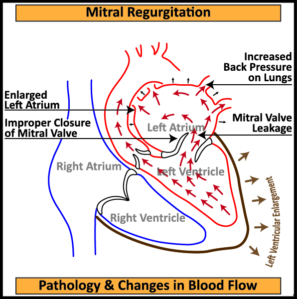
- Regurgitation: In certain diseases, leaflets of Aortic and/or Mitral Valve do not close completely leading to leakage and blood flowing back into the previous chamber (Left Atrium and/or Left Ventricle).
- Aortic Regurgitation: As the disease progresses or due to infection of the leaflets, the Aortic valve may lose their ability to close completely. This results in blood flowing back into the heart from the Aorta leading to increased back-pressure in the heart and lungs causing Palpitation and Breathlessness
- Mitral Regurgitation: As the disease progresses or due to infection of the leaflets, Mitral Valve lose its ability to close completely. This results in blood flowing back into the Left Atrium leading to increased back-pressure in the heart and lungs causing Palpitation and Breathlessness.
Treatment:
The usual investigations needed to diagnose and plan treatment for Double Valve Replacement are ECG, 2D Echo and occasionally, Transesophageal Echocardiography (TEE). In patients above 40 years of age, Coronary Angiography is required to identify Coronary Artery Blockages.
Double Valve Replacement (DVR) Surgery is an Open Heart Surgery. The chest bone (Sternum) is split vertically to access the heart. The diseased Aortic and Mitral Valves are replaced by an artificial (Prosthetic) valve.
The 2 types of Prosthetic Valves available are:
- Mechanical Valve: Made from a composite of Pyrolitic Carbon and Titanium, the Mechanical Valve is generally recommended for younger patients below 60 years of age. The Mechanical Valve is durable and lasts longer. As the material used in construction is inorganic, the patient has to consume blood thinners (Anti Coagulants) all through their life as well as regular blood testing (PT/INR), sometimes as frequent as monthly, to assess the thinness (Anti-Coagulation) status.

- Bio-Prosthetic (Tissue) Valve: The Bio-Prosthetic Valve is made up of biological tissue, usually of Bovine or Porcine origin. This valve is recommended for patients ideally above 65 years of age. The Bio-Prosthetic Valve, which as per manufacturers claim lasts 15 to 18 years, is much lesser than Mechanical Valve. Incase of degeneration of Bio-Prosthetic Valve, patient has to again undergo one or both Valve Replacement Surgery (Redo Surgery). At the same time, patient has to consume blood thinners (Anti Coagulants) for just 3 months after surgery.

Salient Features of Double Valve Replacement Surgery
Irrespective of the type of Prosthetic Valves used, the patient is able to return to an active and healthy life after Double Valve Replacement Surgery.
- The patients display significant symptomatic improvement after Double Valve Replacement Surgery.
- In some cases, where Coronary Artery blockages are observed along with Double Valve malfunctions, both Coronary Artery Bypass Grafting (CABG) & Double Valve Replacement (DVR) Surgeries can be performed simultaneously.
- In some cases, the Aortic & Mitral Valves are damaged along with severe Tricuspid Regurgitation. In such cases, Tricuspid Valve Repair/ Annuloplasty (TV Repair/TVA) Surgery can be performed along with Double Valve Replacement (DVR) Surgery simultaneously.