Pathology:
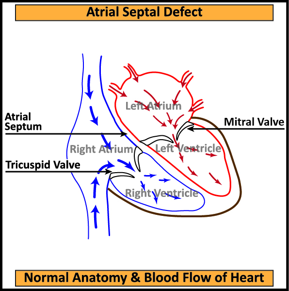
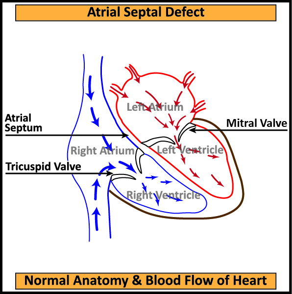
The two upper chambers of the heart, ie Left and Right Atrium are separated by a wall called Atrial Septum. The right chambers of the heart pump oxygen depleted blood from the body to the Lungs. The left chambers receive oxygen rich blood from the lungs and pump it to the body. In some cases, after birth, Atrial Septum does not close fully, leaving a “Hole” between the Left and Right Atrium called Atrial Septal Defect (ASD).
If the hole is small, the patient does not experience any significant symptoms. In patients with Atrial Septal Defect, blood leaks across the hole from Left into Right Atrium. This recirculation of excess blood causes enlargement of the right sided chambers. The excess blood causes enlargement of the right sided chambers. The excess blood also increases pressure in arteries of the lungs, which over a period of time may cause irreversible damage of Lungs (Eisen Menger Syndrome). Atrial Septal Disease can present with Shortness of Breath on Exercise, Fatigue, Palpitation and occasionally Stroke.

Complications:
The disease presents itself in the patient during childhood and occasionally in adulthood. The usual investigations needed to diagnose and plan treatment for Atrial Septal Disease are 2D Echo and occasionally Transesophageal Echocardiography (TEE). In patients above 40 years of age, Coronary Angiography is required to identify Coronary Artery Blockages.
Treatment:
The two types of corrective procedures for Atrial Septal Disease Closure are
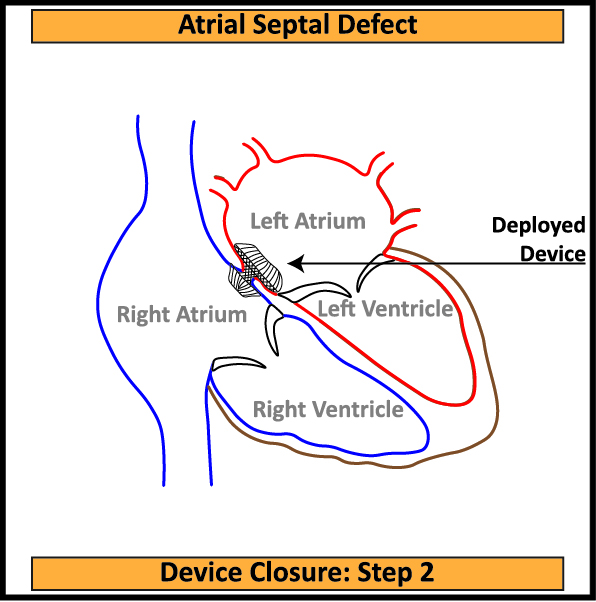
Device Closure (Endovascular Repair): This procedure is performed in a Cathlab under fluoroscopic guidance. A dumbbell shaped device is inserted in the femoral vein via a catheter. The device acts as a plug across the hole in the Septal Wall. Since the plug needs a firm support all around, the procedure cannot be performed if the hole is located near the periphery of the Septal Wall. Device Closure is performed only in small and medium sized holes. The device may dislodge and migrate causing life threatening complications which require emergency surgical intervention.

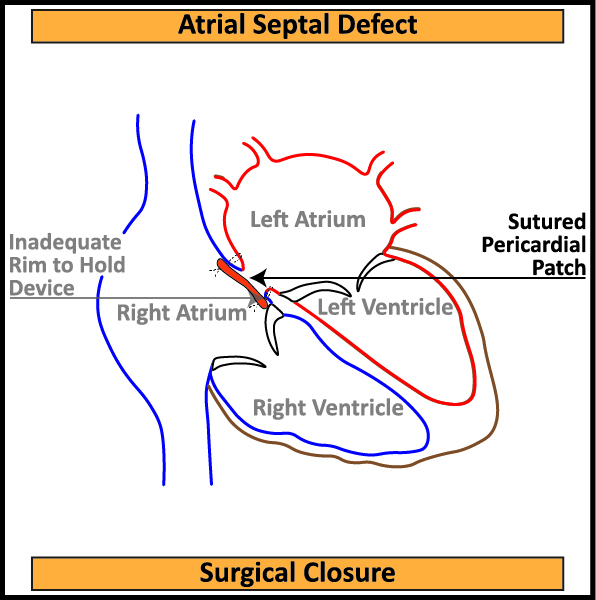
Surgical Correction (Pericardial Patch Closure): In this procedure, a patch is harvested from the Pericardium, which is a natural covering of the Heart. The patch is used to seal the leak between the left and right atria. The two approaches to surgical correction are:
Open Surgical ASD Closure: The chest bone (Sternum) is split vertically to access the heart.
Minimally Invasive ASD Closure (MICS): A small incision is made on the right side of the rib cage to access the heart. The procedure required special instruments and techniques.

Salient feature of Atrial Septal Defect (ASD) Closure Surgery
After surgical correction of Atrial Septal Defect, the patient is able to return to an active and healthy life. The patient can expect to live his normally expected life span without further complications.
Patients display significant symptomatic improvement after Atrial Septal Defect Correction Surgery.
In some cases, where Coronary Artery blockages are observed along with Atrial Septal Defect Closure, both Coronary Artery Bypass Grafting (CABG) & Atrial Septal Defect (ASD) Closure Surgeries can be performed simultaneously.


